The maternity unit at University Hospital of Wales (UHW), Cardiff, UK, adopts the latest technology to enhance routine antenatal screening of fetal heart rate.
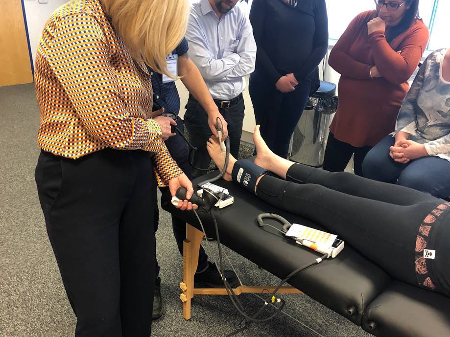
Senior midwifery staff at the hospital have been working with local industry to develop a new handheld fetal Doppler which, in addition to the conventional numeric display of fetal heart rate, now has a trace view screen which it was felt would add value in enhancing the process of intermittent auscultation, and in allowing the procedure to be documented electronically in the maternity record.
The hospital has now invested in a number of these new Dopplers and the value of the trace view was highlighted in a recent case at the hospital.
It was being used routinely to auscultate the fetus in the normal numeric display mode, with a baseline rate of 130bpm being documented. They switched to the trace view and this was reported as showing good variability and the presence of accelerations. A “stretch & sweep” was performed with auscultation being repeated afterwards. The Doppler was still in trace mode and this showed a baseline rate of 100 with noticeable shallow decelerations and reduced variability. This can be normal after such a procedure, but in this case there was sufficient concern that they kept the trace view running until they were able to transfer mum onto a CTG – this showed a bradycardia, triggering a grade 1 emergency C-section, with the baby being delivered within 21 minutes. The baby spent a short time on NICU but made a full recovery.
The midwife involved believed that this new trace view feature alerted her to this situation earlier than would have been the case with a traditional Doppler, enabling her to take appropriate action resulting in a positive outcome.
It is important to note that this trace view is not a substitute for CTG, but it does provide a more robust picture than a simple single “baseline” rate. It can give an indication of baseline rate and an indication of other trace features including variability, accelerations & decelerations, as witnessed by this case. However, there is not enough resolution to measure these details and where any concern arises about fetal condition from using this trace view mode, a full CTG should always be used to confirm this. The trace view can also show maternally sensed fetal movements and this trace record can be stored and transferred to the patient’s electronic maternity record to document the auscultation procedure – another benefit of the trace view, as just storing a random heart rate value to document this would be of little clinical significance.
Disclaimer
All information is provided in good faith – without warranty of any kind, express or implied – and any use of information or material contained herein is at the users’ sole risk. This blog is for information purposes only and shall not be construed as the giving of advice or making of any recommendation and the information should not be relied upon as the basis for any decision or action. The views and opinions of authors expressed herein do not necessarily state or reflect those of Arjo.